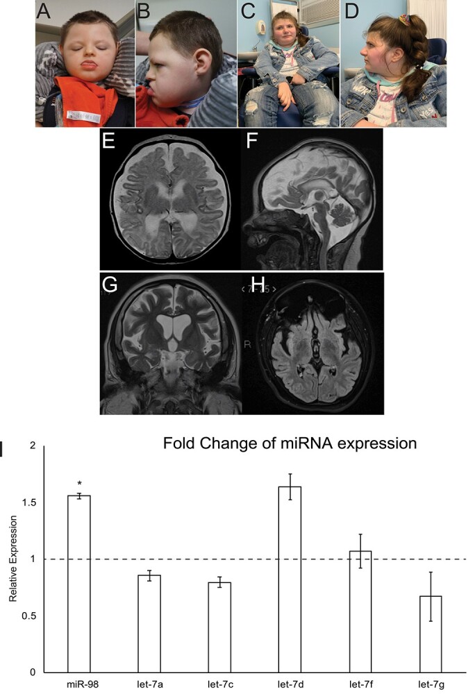
Figure 1.

Two individuals with DROSHA variants show facial dysmorphia, microcephaly and white matter atrophy. (A and B) Individual 1 at 3 years, 11 months. (C and D) Individual 2 at 23 years. (E and F) Axial (E) and sagittal (F) T2 weighted MR images from Individual 1 obtained at age 5 weeks show global atrophy. (G and H) Coronal T2 (G) and axial FLAIR (H) MR images from patient 2 obtained at age 17 years show global atrophy. (I) Expression of a panel of miRNAs in DROSHAD1219G fibroblasts. miRNA expression was assayed using TaqMan assays and compared to control fibroblasts. miR98 expression was significantly upregulated in DROSHAD1219G fibroblasts. *P < 0.5.
