
Figure 3.
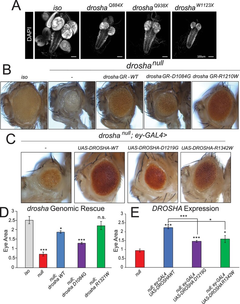
DROSHA variants damage protein function and are partially able to rescue fly eye/head size defects. (A) Third instar larval brains stained with DAPI to show nuclei. Control fly larvae (first panel) display a wild-type brain and attached imaginal discs (first panel). Drosha null allele mutants show dramatically reduced brains as well as a loss of the attached imaginal discs (second–fourth panels). The VNC that can be seen posterior to the two brain lobes is unaffected. (B) Eye-specific droshaW1123X clones were generated using the ey-GAL4 UAS-FLP/FRT system (74). A fly with clones of the isogenized FRT42D chromosome (control, first panel) displays wild-type eye size. droshaW1123X (referred to as droshanull in the images for simplicity, second panel) clones cause both the eye and head size to be reduced. Introduction of a wild-type GR (drosha GR-WT, third panel) construct is able to partially rescue eye and head size defect. Introduction of a GR construct carrying a mutation that corresponds to Individual 1’s variant (drosha GR-D1084G, fourth panel) has about half of the activity of the wild-type construct in this assay, whereas a GR construct carrying the mutation that corresponds to Individual 2’s variant (drosha GRR1210W, fifth panel) has activity that is similar to the wild-type construct. Results are quantified in (D). (C) Expression of DROSHA in the drosha mutant eye clones. Droshanull mutant clones (first panel) have reduced eye size as was shown in (A) and Figure 2D. Expression of wild-type DROSHA (second panel) partially rescues this eye size defect, whereas expression of p.D1219G variant (third panel) again only has about half of the activity of the reference protein. Expression of p.R1342W variant rescues eye size to 75% of the effect of the reference protein (fifth panel). Results are quantified in (E). *P < 0.05,**P < 0.001, ***P < 0.0001.