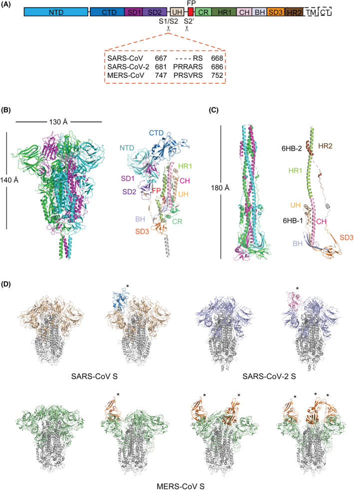
Fig. 1.

Overall structures of the S glycoprotein in the pre‐fusion and post‐fusion states. (A) Schematic representation of the structural domains in the S protomer. These domains are shown as boxes with the width reflecting the relative length of the amino acid sequence. The S1/S2 and S2’ cleavage sites are indicated by scissors. The S1/S2 cleavage sites of SARS‐CoV, SARS‐CoV‐2 and MERS‐CoV are shown in the dashed orange box. NTD, N‐terminal domain; CTD, C‐terminal domain; SD1, subdomain 1; SD2, subdomain 2; UH, upstream helix; FP, fusion peptide; CR, connecting region; HR1, heptad repeat 1; CH, central helix; BH, β‐hairpin; SD3, subdomain 3; TM, transmembrane domain; CT, cytoplasmic domain. (B) Overall structure of the SARS‐CoV‐2 S glycoprotein in the pre‐fusion state (PDB code: 6XR8). Left: Three protomers are shown as cartoon representations and coloured magenta, green and blue, respectively. Right: Structural domains of a protomer are coloured and labelled according to panel A. (C) Overall structure of the SARS‐CoV‐2 S glycoprotein in the post‐fusion state (PDB code: 6XRA). Left: Three protomers are shown as cartoon representations and coloured magenta, green and blue, respectively. Right: Structural domains of a protomer are coloured and labelled according to panel A. 6HB‐1, six‐helix bundle 1; 6HB‐2, six‐helix bundle 2. (D) Structural heterogeneity of SARS‐CoV, SARS‐CoV‐2 and MERS‐CoV S trimers. All S2 subunits are coloured grey. S1 subunits are coloured wheat for SARS‐CoV, light blue for SARS‐CoV‐2 and green for MERS‐CoV. ‘Up’ RBDs are indicated by asterisks and coloured blue for SARS‐CoV, pink for SARS‐CoV‐2 and orange for MERS‐CoV. SARS‐CoV S (PDB: 6ACC and 6ACD); SARS‐CoV‐2 S (PDB: 6VXX and 6VYB); MERS‐CoV S (PDB: 6Q05, 5X5F, 5X5C and 5X5C). [Colour figure can be viewed at wileyonlinelibrary.com]
