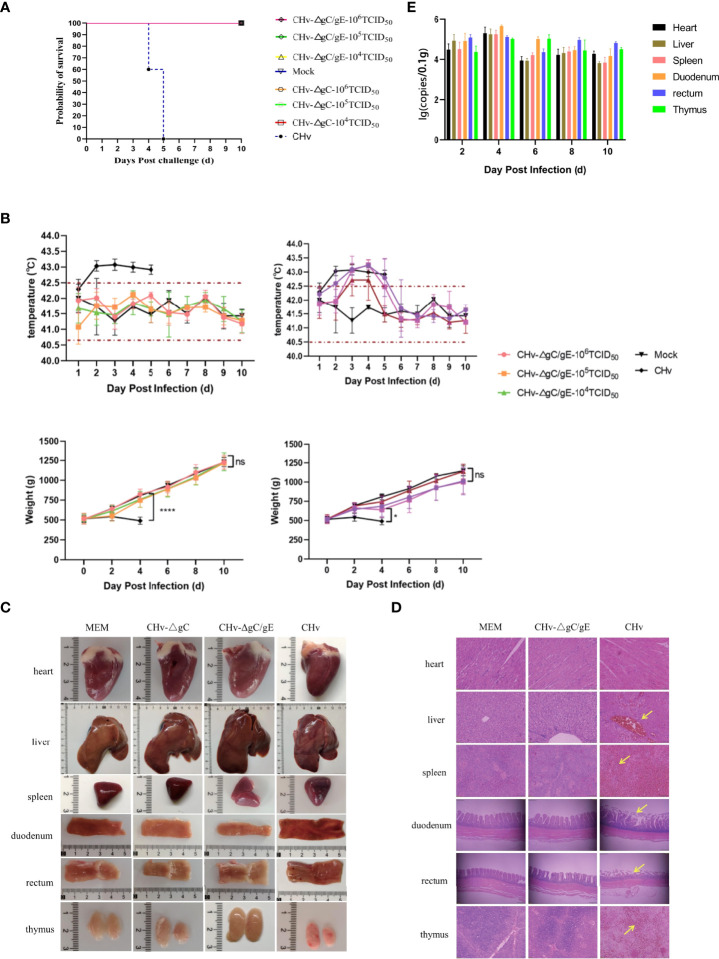
Figure 4.
Pathogenicity of CHv-ΔgC/gE in ducklings. (A) Survival curves of ducklings after challenge with the indicated viral strains. (B) Daily body weight and temperature of all the ducklings after challenge with the indicated viral strains. (C) Postmortem examination of ducklings in each group at 5 dpi. (D) Histological analysis of heart, liver, spleen, duodenum, rectum and thymus from ducks injected with the indicated viral strains or MEM. Arrows indicate cellular infiltration or tissue disruption. (hematoxylin and eosin staining, 100× magnification). (E) Quantification of viral DNA loads in selected tissues with real-time PCR. Viral DNA copy numbers were determined with primers specific for UL30, as described above. *p<0.05; **p<0.01; ***p<0.001; ****p<0.0001; and ns, not significant.

