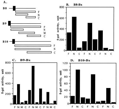
FIG. 3.
Delineation of interaction domains of VirB8, VirB9, and VirB10. Interactions of VirB8 (B), VirB9 (C), VirB10 (D), and their derivatives with VirB8, VirB9, and VirB10 were investigated by the two-hybrid assay. (A) A linear representation of the VirB proteins and their derivatives used for fusion construction. Fusions containing the periplasmic domain (F), an N-terminal fragment (N), a central fragment (M), and a C-terminal fragment (C) were tested for interaction with the three proteins. The solid box indicates a hydrophobic region.

