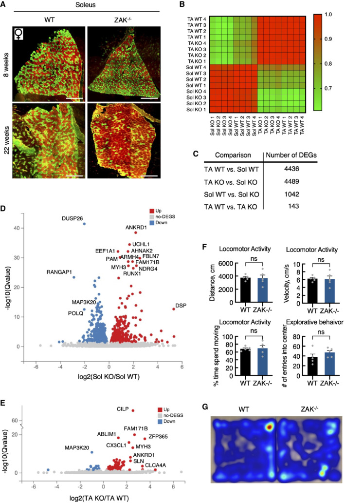
Figure EV5. ZAK−/− mice present with muscle pathology.

-
ASoleus muscle cross‐sections from 8‐ and 22‐week‐old WT and ZAK−/− female mice were immunostained for type I (red) and type IIa (green) fibers using myosin isoform‐specific antibodies. Scale bars, 500 μm.
-
BCorrelation map of transcriptomes from all individual samples color‐coded according to the correlation coefficient.
-
CNumber of differentially expressed genes (DEG) for the indicated group comparisons.
-
DVolcano plot of up‐ and downregulated DEGs in soleus muscle dependent on genotype.
-
EVolcano plot of up‐ and downregulated DEGs in TA.
-
FGeneral locomotor activity of 16‐18‐week‐old WT and ZAK−/− male mice was evaluated using a standard open field test. No difference in distance traveled, velocity, time spent moving, or number of entries to the center zone was observed between the genotypes. Error bars represent the standard deviations (n = 5 biological replicates). ns—not significant in unpaired t‐test.
-
GHeat maps illustrating representative moving patterns. TA—tibialis anterior; Sol—soleus.
