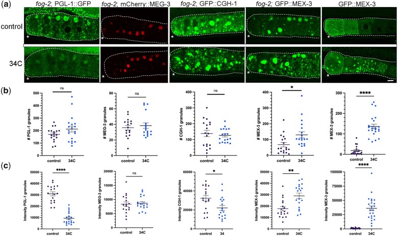
Fig. 1.
Increased temperature induces de-condensation and condensation of RNA-binding proteins. a) Micrographs of 4 fluorescently tagged RNA-binding proteins in a fog-2 background, and GFP::MEX-3 in diakinesis oocytes. Top row: arrested oocytes under control conditions of 20°C. Bottom row: arrested oocytes after exposure to 34°C for 40 min (PGL-1) or 2 hr (all other proteins). Oocytes are outlined in white dashed lines. Asterisk marks the most proximal oocyte in each germ line. Scale bar is 10 µm. b) Graphs showing the number of GFP granules in a single Z-slice of proximal oocytes (see Methods). For (b) and (c), statistical significance was determined using the Mann–Whitney test. **** indicates P < 0.0001, ** indicates P < 0.01, * indicates P < 0.05, and ns indicates not significant. n = 17–23. Error bars indicate mean ± SEM. c) Graphs showing the total fluorescence intensity within granules in proximal oocytes (see Methods).

