Fig. 5.
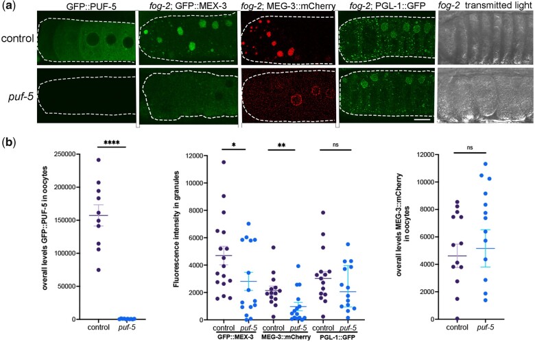
PUF-5 may promote condensation of MEX-3 and MEG-3 into large granules. a) Micrographs of GFP::PUF-5 in diakinesis oocytes, 3 fluorescently tagged RNA-binding proteins in a fog-2 background, and fog-2 oocytes visualized by transmitted light. Depletion of control lacZ or puf-5 was performed by feeding RNAi. The GFP::PUF-5 strain was used as a positive control in each experiment to assess the extent of depletion of puf-5. The GFP::MEX-3, MEG-3::mCherry, and PGL-1::GFP reporters were used to assay the condensation of the respective proteins in arrested oocytes. Oocytes are outlined in white dashed lines, with the most proximal oocyte on the left. Scale bar is 10 µm. b) Graphs showing the overall levels of GFP::PUF-5 or MEG-3::mCherry in oocytes (CTCF, see Methods), or the intensity of fluorescence in granules, as plotted on the Y-axes. Statistical significance was determined using the Mann–Whitney test. **** indicates P < 0.0001, ** indicates P < 0.01, * indicates P < 0.05, and ns indicates not significant. n = 10–22. Error bars indicate mean ± SEM.

