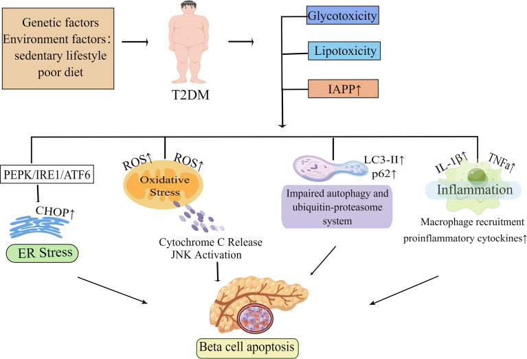
Figure 1.
Molecular mechanisms of beta-cell apoptosis under conditions of IAPP aggregation and glucolipotoxicity. In obesity-associated type 2 diabetes, elevated islet amyloid polypeptide (IAPP), glucotoxicity, lipotoxicity and glucolipotoxicity are the most studied causative factors of beta-cell apoptosis. These factors activate all ER stress pathways in beta-cells, namely the PKR-like ER kinase (PERK), inositol-requiring enzyme 1 (IRE1), and activating transcription factor 6 (ATF6) pathways, and subsequently induce ER stress. When ER stress is prolonged or excessive, it may mediate beta-cell dysfunction and apoptosis by increasing the expression of the pro-apoptotic factor CHOP. In the presence of these pathogenic factors, it also leads to mitochondrial dysfunction and increases the production of reactive oxygen species (ROS). The increase of ROS activates the apoptotic pathway mediated by oxidative stress and mitochondrial cytochrome C in beta-cell. Autophagy can degrade damaged or misfolded cellular components and proteins under normal physiological conditions. However, under conditions of glucolipotoxicity and increased IAPP, beta-cells are subjected to sustained metabolic stress that leads to impaired autophagy which ultimately exacerbates beta-cell dysfunction thereby leading to apoptosis. Islet inflammation often occurs during T2D development and is characterized by macrophage recruitment to infiltrating immune cells, which can lead to increased production of cytokines and chemokines such as IL-1β, TNFa, and these pro-inflammatory signals can activate apoptotic mechanisms, including ER stress and oxidative stress in beta-cells. By Figdraw (www.figdraw.com).

