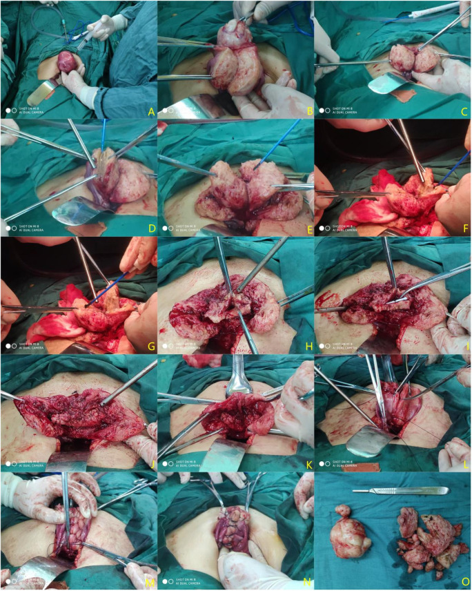
Figure 1.
Procedure diagram of a transabdominal operation. (A) Inject diluted pituitrin; (B) remove uterine fibroids; (C) make a longitudinal incision of adenomyosis lesions to reach the uterine cavity; (D) Resect the lesion as much as possible (preserving approximately 0.5–1 cm of the plasmomuscular layer flaps); (E) treat the contralateral lesions with the same method; (F) gradually subtract the lesion to the uterine cavity and excise a part of the uterine cavity to reduce the uterine volume; (G) treat the contralateral lesions with the same method; (H) remodel the myometrium; (I) place the LNG-IUS (Manchester ring) and reshape the depth of the uterine cavity again based on the LNG-IUS length; (J) Mattress-suture the uterine cavity continuously; (K) align the sarcoplasmic layers and reduce the extra length; (L,M) suture the bilateral seromuscular layers with the “baseball stitching technique”; (N) repair the sutured uterus; (O) uterine fibroids and adenomyosis specimens resected during the operation.

