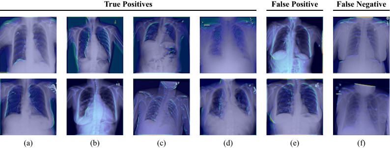
Figure 6.
Grad-CAM visualizations for the first convolutional layer of the ResNet-50 incorporated in our SNN. Each column represents one image pair. The first four columns (a–d) show true positive classifications. The last two columns (e) and (f) depict a false positive and a false negative classification, respectively. The shown images illustrate that the anatomical structure of, e.g. the breast (cf. (a,b,e)), the lungs (cf. (a,b,c,e)), and the heart (cf. (a,b)) have a high impact on the final model prediction. Furthermore, it can be seen that our network focuses on the collarbones (cf. (a,c,d,f)) and the ribs (cf. (b,c,e)). The upper images of (a) and (f) also highlight that our network pays attention to the contour of the diaphragm.

