Figure 6.
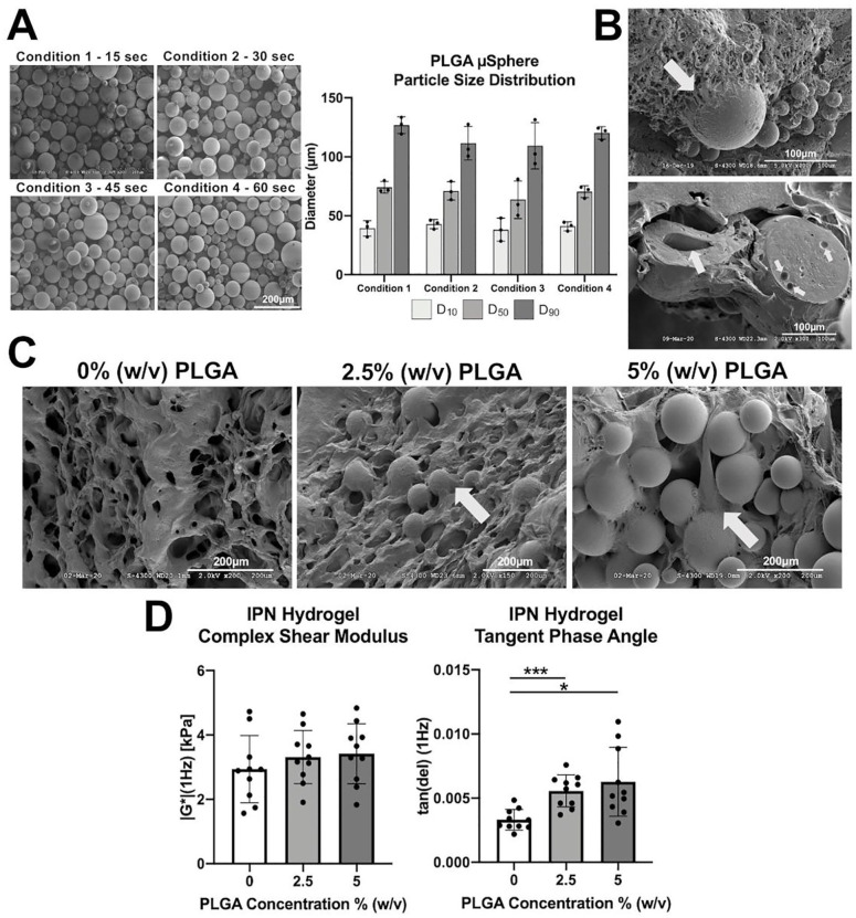
PLGA µSpheres integrate within the IPN hydrogel’s bioadhesive polymer network, do not significantly increase the complex shear stiffness upon hydrogel embedment, and significantly increase the energy dissipation potential of the IPN hydrogel after equilibrium swelling. (A) SEM and laser diffraction analysis show no difference in µSphere morphology or particle size distribution, respectively, when minimizing vortexing time in the double emulsion fabrication protocol. D10 = 10th percentile PLGA µSphere diameter, D50 = 50th percentile PLGA µSphere diameter, and D90 = 90th percentile PLGA µSphere diameter. Panel A scale bar = 200 µm. (B) SEM demonstrating hydrogel polymerization around the µSphere surface upon embedment (white arrows—top image) and that µSpheres are mononuclear and polynuclear by composition (white arrows—bottom image). Panel B scale bar = 100 µm. (C) Microstructure of the PLGA µSphere delivery system within the IPN hydrogel for 2.5% (w/v) and 5% (w/v) conditions compared with µSphere-free controls (0% [w/v]). Panel C scale bar = 200 µm. (D) Complex shear stiffness (|G*|) and tangent phase angle (tan δ) at 1 Hz for IPN hydrogels with 0% (w/v), 2.5% (w/v), and 5% (w/v) PLGA µSpheres after 72 hours of equilibrium swelling in 1X phosphate-buffered saline. PLGA = poly(lactic-co-glycolic acid); IPN = interpenetrating network; SEM = Scanning Electron Microscopy. *P < 0.05; ***P < 0.0005.

