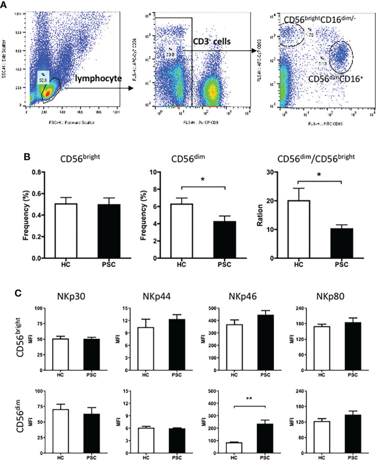
Figure 1.

Characteristics of peripheral NK cells in patients with PSC. PBMCs from PSC and HCs were isolated and stained with fluorochrome-conjugated antibodies for cell subsets. (A) Strategy of FCM of NK cell subsets. CD3 and CD56 expression in PBMCs was analyzed after application of a lymphocyte gate. CD3-negative cells were further gated for analysis of CD3−CD56brightCD16− (CD56bright) and CD3−CD56dimCD16− (CD56dim) NK cell subsets. (B) The frequency (%) of CD56bright and CD56dim cells in total lymphocytes and (C) MFI of natural cytotoxicity receptors in each NK subset were compared between PSC (n = 34) and HCs (n = 29). *, p < 0.05; **, p < 0.01 (two-tailed Mann–Whitney test). NK, natural killer; PSC, primary sclerosing cholangitis; PBMCs, peripheral blood mononuclear cells; HCs, healthy controls; FCM, flow cytometry; MFI, mean fluorescence intensity.
