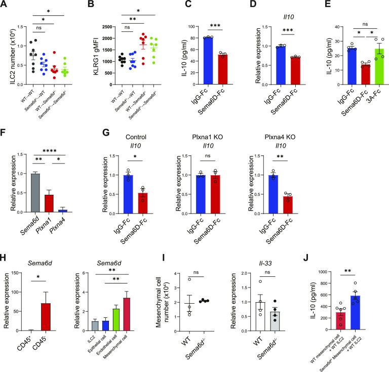
Figure 4. Sema6D signaling from tissue niches suppresses IL-10–producing ILC2s.
(A, B) Lung ILC2 numbers and KLRG1 expression in BM chimeric mice of the indicated genotypes. (C, D, E) Lung ILC2s from WT mice were cultured with Sema6D-Fc, Sema3A-Fc, or IgG-Fc (10 nM each) and stimulated with IL-2 and IL-33 (10 ng/ml each) for 3 d. (C) The amount of IL-10 in the supernatant was measured by ELISA. (D) The mRNA expression of Il10 in (C) was evaluated by qRT-PCR. (E) The amount of IL-10 in the supernatant was measured by ELISA. (F) The mRNA expression of Sema6d, Plxna1, and Plxna4 in WT lung ILC2s was evaluated by qRT-PCR. (G) Freshly sorted ILC2s were prestimulated with IL-2 and IL-33 on day 0 and infected with lentiviral vector (Plxna1 KO, Plxna4 KO, or control vector [mock]) on day 1 and 2. Puromycin-resistant cells were sorted as infected cells on day 7 and stimulated with IL-2 and IL-33 for 72 h. The mRNA expression of Il10 in ILC2s was evaluated by qRT-PCR. (H) qRT-PCR analysis of the Sema6d mRNA in lung cells isolated from WT mice at steady state. (I) The lung mesenchymal cell number and Il-33 mRNA expression were evaluated by qRT-PCR. (J) Concentrations of IL-10 in the supernatants of WT lung ILC2s after 3 d of co-culture with lung mesenchymal cells. Data are representative of two independent experiments (mean ± SEM). *P < 0.05; **P < 0.01; ***P < 0.001 by one-way ANOVA with Tukey–Kramer test (A, B, E, F, H) or t test (C, D, E, G, H, I, J).

