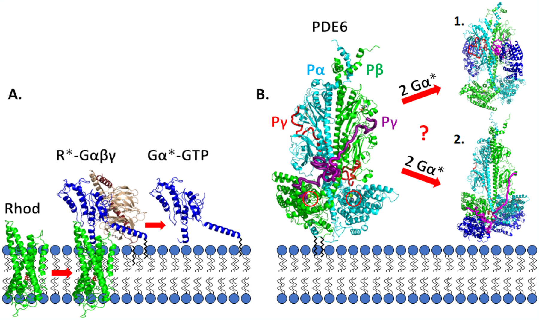
Fig. 1.

Schematic diagram of the visual excitation pathway occurring on the disk membrane of rod photoreceptors A. Photoisomerization of 11-cis retinal to all-trans retinal (red) bound to the visual receptor rhodopsin (Rhod, green) induces conformational changes in rhodopsin (R*). Enhanced binding of R* to the photoreceptor G-protein, transducin (Gαβγ), results in GTP exchange on the G-protein α-subunit (Gα, blue) and dissociation of Gα*-GTP from R* and from Gβγ (tan and brown). B. Rod PDE6 holoenzyme (αβγγ) is maximally activated upon binding of two Gα*-GTP molecules that result in displacement of the intrinsically disordered Pγ subunits from the enzyme active site (red circles). Two different mechanisms of Gα* activation of PDE6 [50,51] are discussed in the Section 5.2. Black zig-zag lines represent post-translational modifications: Gα, heterogeneous N-terminal acylation [135,86]; Gγ, farnesyl group [136,137]; Pαβ, farnesyl and geranylgeranyl groups [138]. Space-filling models were generated from the following sources: R*-Gαβγ [139]; Gα*-PDE6 activated complexes [50,51].
