
Fig. 6.
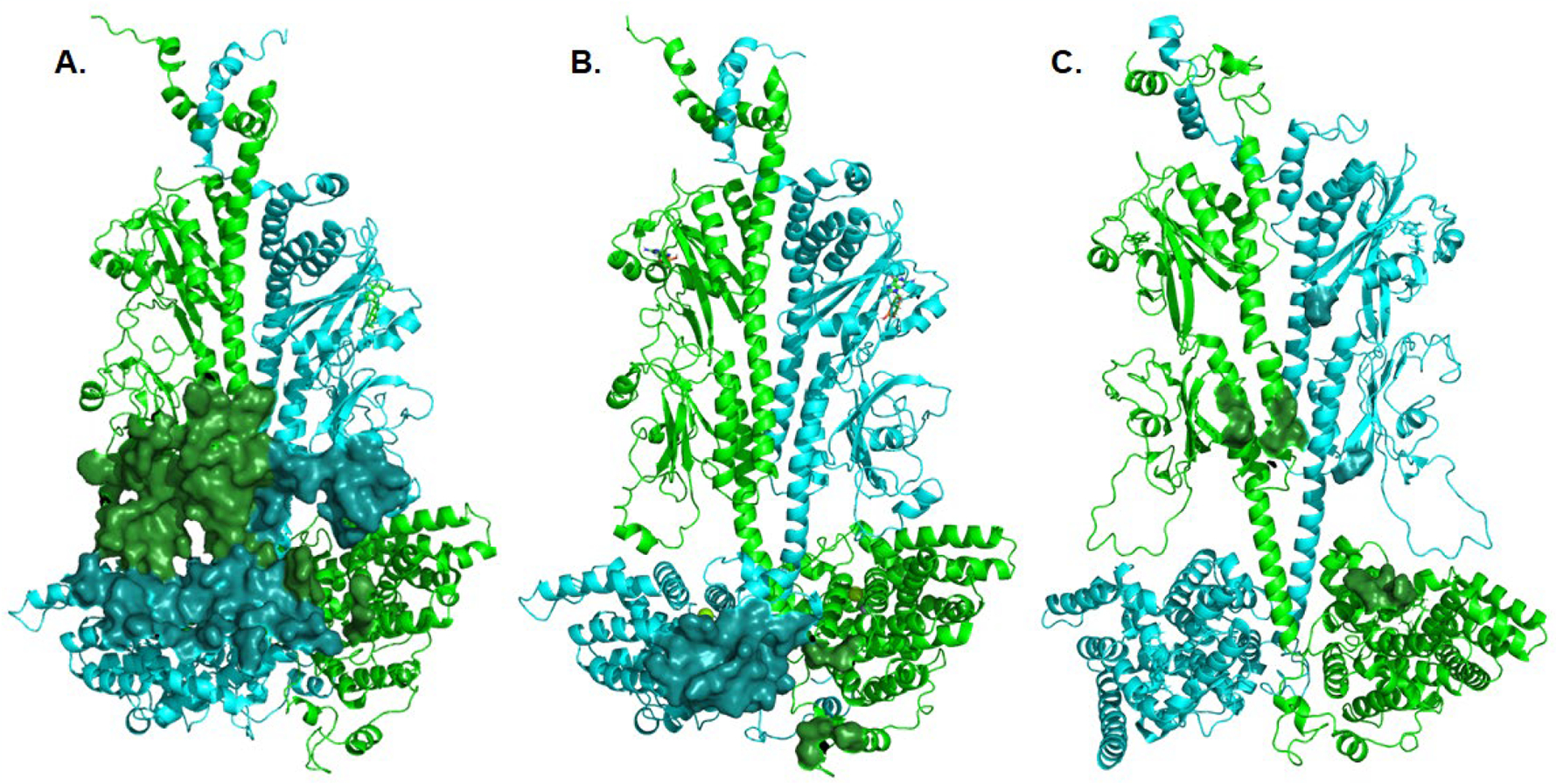
Structural models of the interacting residues between Gα* and Pαβ Interface residues between Pα (cyan) and Pβ (green) with the Gα* subunit were calculated using the Interface Residues script in Pymol, and depicted as a solid surface. A. Interactions of a Gα* subunit with the GAFb domain of PDE6 (derived from Fig. 3C of [50]). B. Interactions of a Gα* subunit with the catalytic domain of PDE6 (derived from Fig. 3A of [50]). C. Interactions of a chimeric Giα/Gα* with the GAFb domain of PDE6 (derived from PDBID: 7jsn; [51]). For clarity, the Gα and Pγ subunits are not shown.