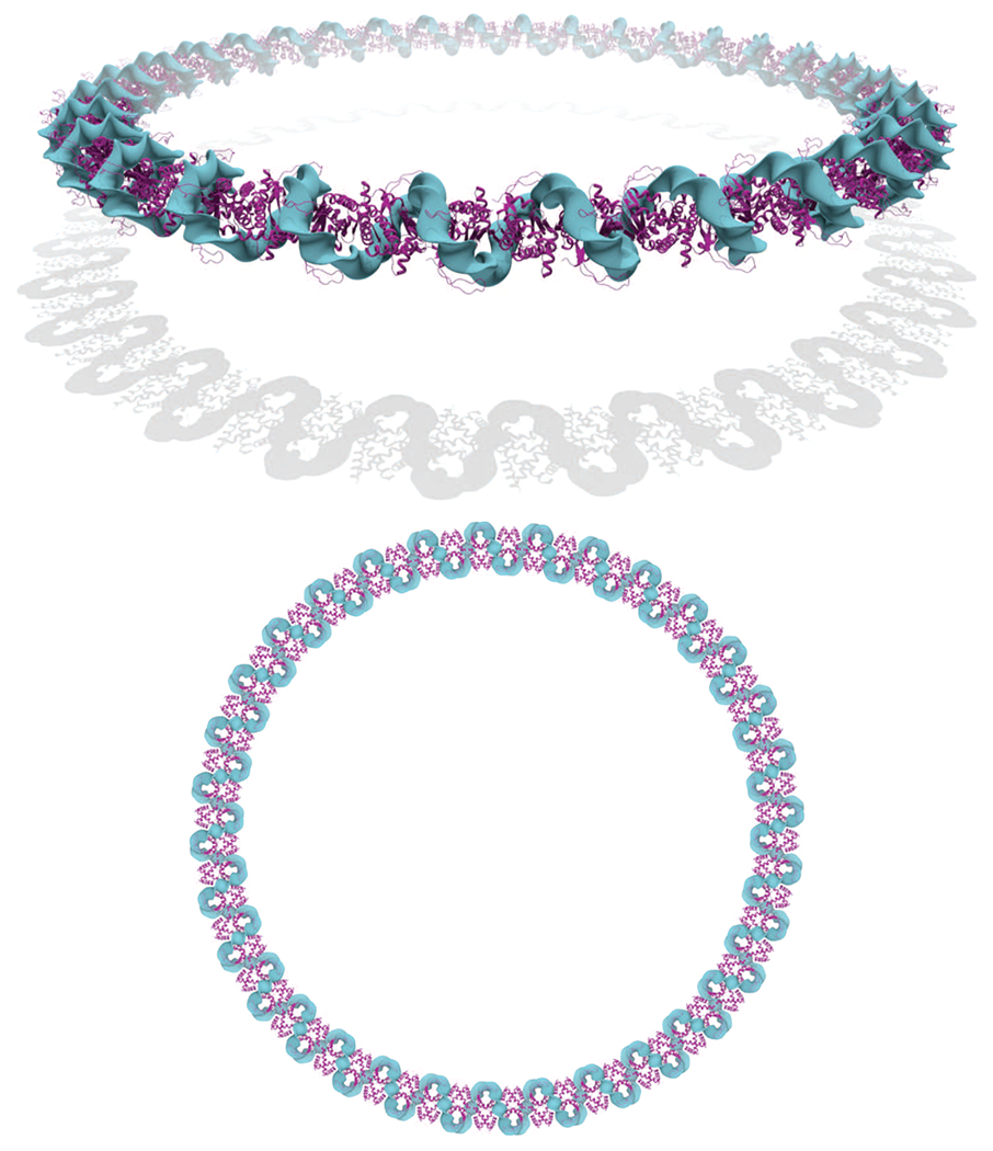
Figure 5.

Perspective (top) and orthographic (bottom) views of an optimized DNA plasmid of 1280 bp containing 64 Hbb proteins regularly spaced by 5 bp protein-free linkers. The presence of the Hbb proteins (represented in magenta) greatly condenses the DNA into a helical fiber-like structure that folds into a circular configuration. The circular radius of the resulting superstructure is 31.5 nm, and the fiber radius is 3 nm (the same DNA plasmid without any proteins has a circular radius of 69.2 nm and a cross-sectional radius of 1 nm). The elastic energy stored in each naked-DNA linker is 0.19 kBT, suggesting that the linkers are only slightly deformed. In contrast to the zigzag superhelical structure found in the crystal lattice in which the proteins lie on opposite sides of the superhelical axis,5 the proteins form a central core in the optimized structure. Notice that this structure has been generated with a reduced Hbb model with only the 15 central base pairs of the full crystallographic model, i.e., with 10 bp peeled off each end of the protein-bound DNA fragment used in earlier figures. As a consequence, the spacing between the proteins is smaller in the optimized structure than in the pseudocontinuous helix found in the crystal lattice.
