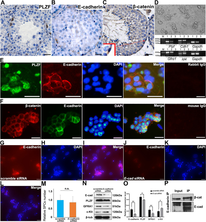
Fig. 1.
Expression of E-cadherin and SPC markers in mouse testis and isolated SPCs. The expression of PLZF (A), E-cadherin (B) and β-catenin (C) was detected in the testis of 42-day old mouse using IHC. The morphology of purified SPCs on MEF feeder layer was exhibited, and expression of SPCs markers was determined using RT-PCR (1.testis, 2.SPCs, 3.H2O) (D). Co-IF staining was employed to detect the expression of PLZF and E-cadherin (E green: PLZF, red: E-cadherin, blue: DAPI), or β-catenin and E-cadherin (F red β-catenin, green E-cadherin, blue DAPI) in purified SPCs. Expression of E-cadherin was detected in SPCs transfected with scrambled (G E-cadherin, H. DAPI, I. Merge) or E-cadherin siRNA (J E-cadherin, K DAPI, L Merge) was exhibited 72 h post transfection using IF staining. The number of SPCs was statistically calculated (M) (10 views of ×200 were randomly selected). The expression of E-cadherin, PLZF, GFRA1, c-Kit and β-tubulin was determined in scrambled or E-cadherin siRNA transfected SPCs using Western blot, n=3 (N), and was statistically calculated (O). The interaction of E-cadherin and β-catenin in SPCs was detected using co-IP (P). Scale bar = 20 μm, data represents mean ± standard deviation (SD), *p<0.05, **p<0.01

