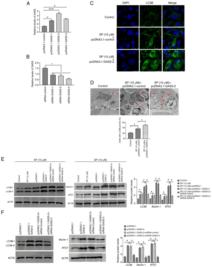
Figure 2.
GAS5 promotes SP-induced autophagy in PAECs. (A) Transfection efficiency of pcDNA3.1-GAS5 in PAECs. (B) Transfection efficiency of siRNA-GAS5 in PAECs. (C) The abundance of LC3B in PAECs treated with the combination of SP (10 µM) and pcDNA3.1-GAS5, as assessed by immunofluorescence assays (scale bar, 25 µm; magnification, ×20). (D) Autophagic vacuoles in the cellular cytoplasm of PAECs treated with the combination of SP (10 µM) and pcDNA3.1-GAS5, as evaluated by transmission electron microscopy (scale bar, 2 µm; magnification, ×10,000). Red arrows indicate the autophagic vacuoles. (E) Protein expressions of LC3B, Beclin-1 and ATG7 in SP (10 µM)-treated PAECs transfected with pcDNA3.1-GAS5 and the combination of pcDNA3.1-GAS5 and siRNA-GAS5, as assessed by Western blotting. (F) Abundance of LC3B in PAECs transfected with pcDNA3.1-GAS5 and the combination of pcDNA3.1-GAS5 and siRNA-GAS5, as assessed by Western blotting. *P<0.05. There were at least three replicates in each group available for analysis. GAS5, growth arrest-specific transcript 5; SP, spermidine; PAECs, pulmonary artery endothelial cells; siRNA, small interfering RNA.

