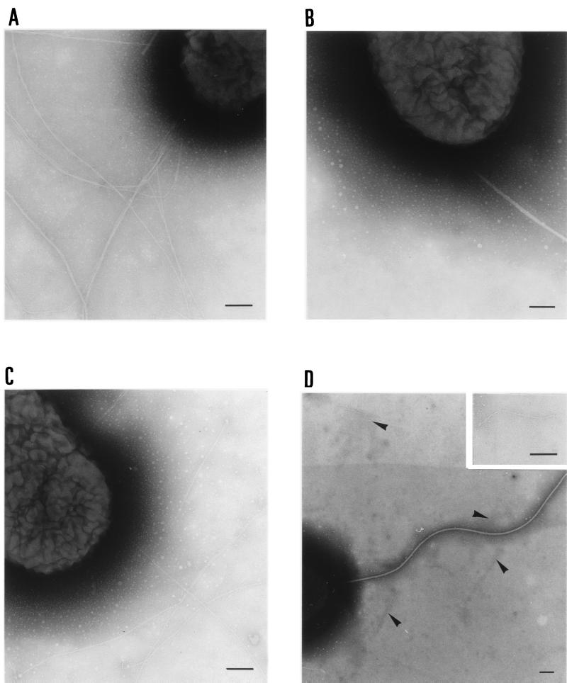
FIG. 3.
Visualization by electron microscopy of pili produced by P. aeruginosa PAK derivatives. Bacteria bearing plasmids carrying cloned genes were grown on medium containing 150 μg of carbenicillin per ml. Basic electron microscopy procedures were as described in the legend to Fig. 2. P. aeruginosa PAK (A), PAK A− (B), PAK A− A+(pAWJ103) (C), and PAK A− PpdD+(pCHAP3117) (D) are shown. The arrows in panel D indicate the short filaments produced by PAK A− PpdD+. The insert in panel D shows a higher-magnification view of a short filament. Bars, 100 nm.

