Fig. 3.
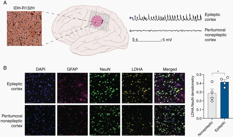
The epileptic human cortex demonstrates upregulation of LDHA compared to the nonepileptic cortex. (A) Diagram demonstrating epileptic cortex (blue electrode) and peritumoral nonepileptic cortex (grey electrode) in the setting of the IDH mutant glioma determined via intracranial EEG monitoring. The glioma stained positive for IDH (R132H) mutation. This patient had a WHO Grade III Astrocytoma IDH-mutant. (B) Multiplex immunofluorescence staining for DAPI (blue), NeuN (green), GFAP (purple), and LDH-A (yellow) of the peritumoral nonepileptic cortex and epileptic cortex demonstrating increased LDH-A expression primarily in neurons. DAPI is a control nuclear DNA stain, GFAP stains astrocytes, NeuN stains neurons, and LDH-A, the metabolic enzyme of interest. LDH-A co-staining with NeuN is significantly increased in the epileptic cortex compared to the peritumoral nonepileptic cortex (n = 4, t(3) = 3.799, P = .0320, paired t-test).

