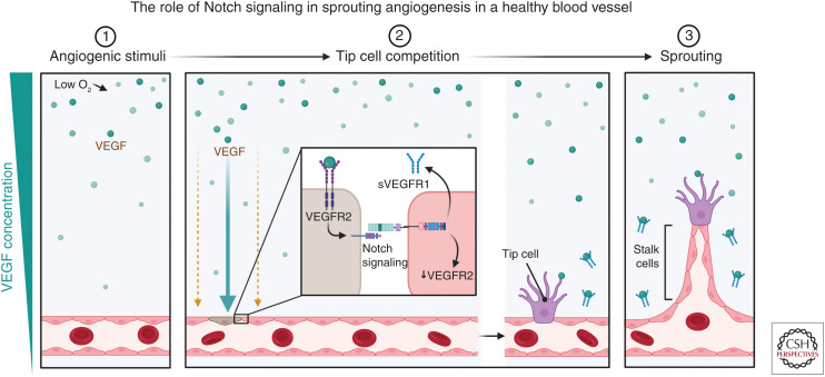
Figure 3.
Schematic of Notch signaling in sprouting angiogenesis. (1) Angiogenesis is initiated by a stimulus such as hypoxia, which leads to an increase in vascular endothelial growth factor (VEGF) expression in tissues. (2) The presence of VEGF (green circles) leads to the binding of VEGF receptor 2 (VEGFR2) on the surface of the endothelial cells. A VEGF/Notch-regulated mechanism ensures a limited number of tip cells are formed through a process known as lateral inhibition. After VEGF binds to the VEGFR2 receptor, it promotes the formation of a tip cell (brown cell) and promotes an increase in the Notch ligand Dll4 expression, while simultaneously inhibiting the formation of tip cells by its neighbors through Notch signaling. Activating Notch signaling results in the down-regulation of VEGFR2 and promotes the production of soluble VEGFR (sVEGFR) that can then act to scavenge extracellular VEGF and hence prevent overvascularization. (3) These cells will then become stalk cells that form the body of the sprouting vessel. (Created in BioRender.com.)

