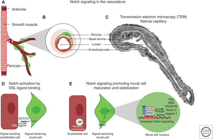
Figure 4.
The role of Notch in vascular endothelial and mural cell communication. (A) Schematic of mural cell localization within branching arterioles. (B) Cross-sectional magnification of a capillary highlight pericytes (green), endothelial cells (red), and basal lamina (pink). (C) Transmission electron microscopy (TEM) images of vessels within the retinal ganglion cell layer demonstrate a peg and socket connection between the endothelial cell and pericyte. (D) Notch expressed in the mural cell (green) is activated upon ligand binding from the endothelial cell (red). (E) After a series of proteolytic events (see Fig. 1), the Notch intracellular domain (NICD) translocates to the nucleus and promotes transcription of a number of genes including PDGFRβ, smooth muscle actin (SMA), Notch 3, and Jagged 1, which promote mural cell maturation and stability. (DSL) Delta/Serrate/Lag, (RBPJK) recombination signal-binding protein for immunoglobulin κJ. (Created in BioRender.com.)

