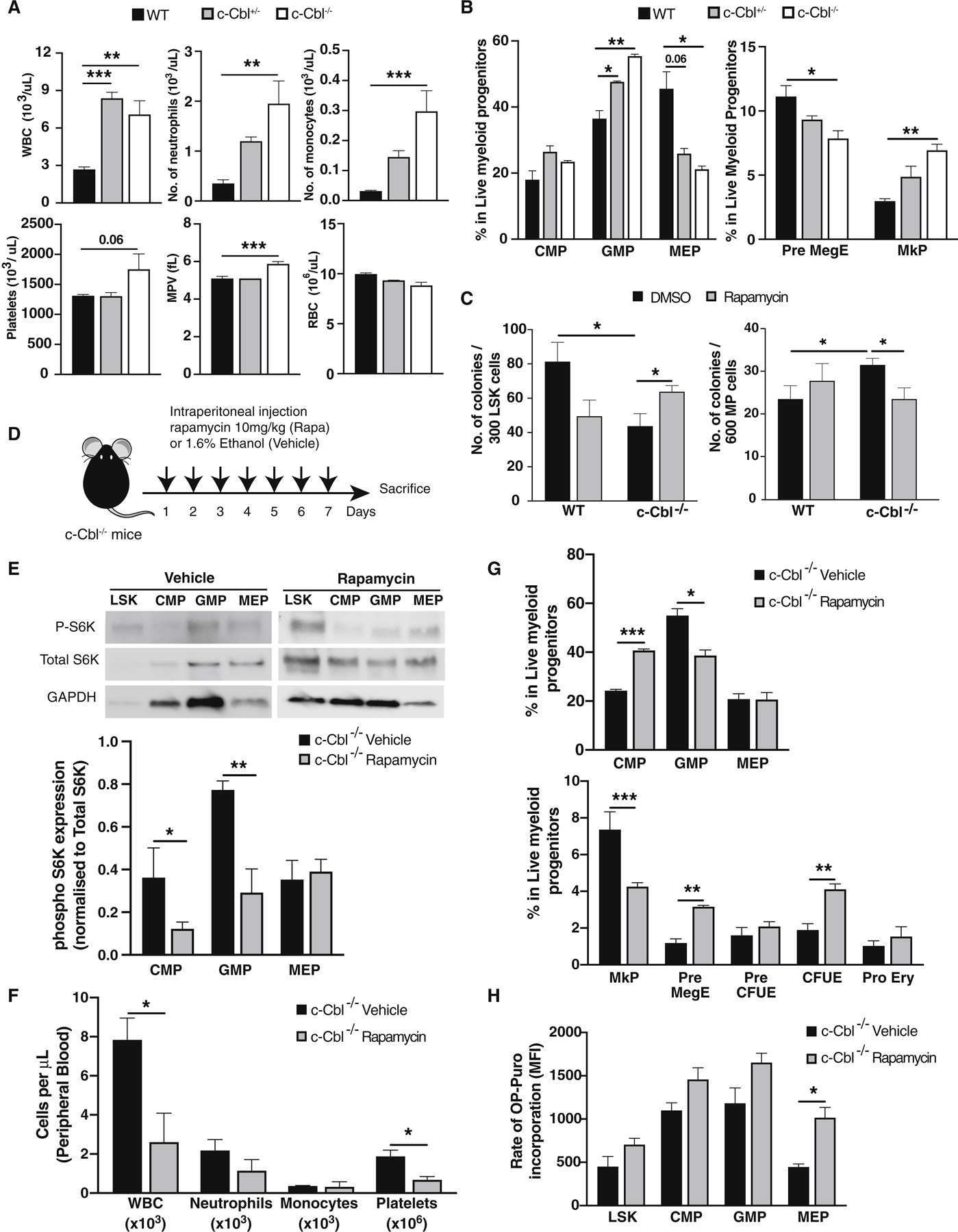
Figure 3. Loss of c-Cbl results in aberrant mTOR signaling in myeloid progenitors and increased mature myeloid cell output.

(A) Complete blood counts from WT, c-Cbl+/− and c-Cbl−/− mice (n=4–5). (B) Flow cytometric analysis of bone marrow cells in the myeloid progenitor (MP) compartment from WT, c-Cbl+/−, and c-Cbl−/− mice. Frequencies were determined using mononuclear cells from two femurs and two tibiae (n = 3–4 mice). (C) Colony forming activity of WT and c-Cbl−/− LSK and MP cells plated in complete methylcellulose for 8 days treated with DMSO or rapamycin (20nM) (n=3). (D) Experimental scheme for in vivo rapamycin treatment studies in c-Cbl−/− mice (n=3/group). (E) Representative immunoblots of mTOR signaling intermediates in LSK and MP subpopulations (CMP, GMP and MEP) from rapamycin treated mice. Phosphorylated p70S6K was quantified by normalizing to both total S6K and GAPDH expression (n=3). (F) Complete blood counts in vehicle and rapamycin treated c-Cbl−/− mice. (G) Flow cytometric analysis of bone marrow MP subpopulations in vehicle and rapamycin treated c-Cbl−/− mice. (H) OP-Puro incorporation assays in LSK and MP cells from vehicle and rapamycin treated c-Cbl−/− mice.
