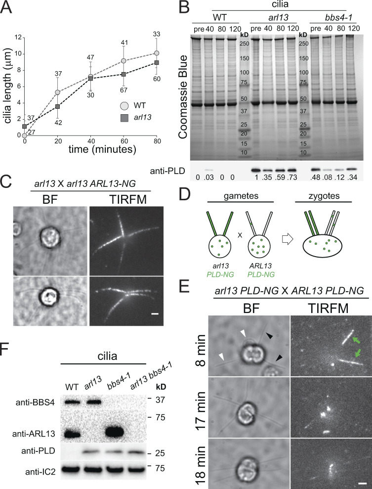
Figure S3.
PLD accumulates slowly in arl13 cilia. (A) Cilia regeneration kinetics after deciliation by a pH shock for control (WT; circles) and arl13 (squares). One cilium was measured per cell, and the number of cells analyzed is indicated. (B) Coomassie Blue stained gel (top) and Western blot of a replicate gel using anti-PLD (bottom) of control (g1, WT), arl13 and bbs4-1 cilia harvested at full-length (pre; no deciliation) and during regeneration at 40, 80, and 120 min after deciliation by a pH shock. PLD accumulated over time in regenerating arl13 and bbs4-1 cilia. Note transient presence of PLD in control cilia during early regeneration. Numbers indicate the anti-PLD signal strength normalized for the Coomassie Blue stained samples. One of two biological replicates is shown with the other experiment omitting the 40 min-samples and the bbs4-1 strain. (C) Bright field (BF) and TIRF images of two arl13 × arl13 ARL-NG zygotes. The images were recorded within 20 min after mixing of the gametes indicating rapid entry of ARL13-NG in arl13-derived cilia. Bar = 2 µm. (D) Schematic presentation of the dikaryon rescue experiment using arl13 PLD-NG and control PLD-NG gametes. After fusion, ARL13 provided by wild-type parent is available for entry into arl13-derived cilia and its effect on the distribution of PLD-NG can be analyzed by TIRFM. (E) Gallery of bright field (BF) and TIRF images of arl13 PLD-NG × PLD-NG zygotes. The time passed since mixing of the gametes is indicated. In the top row, green arrows and black arrowheads indicate the PLD-NG positive cilia; white arrowheads mark cilia derived from the PLD-NG parent. Bar = 2 µm. (F) Western blot of isolated cilia from control (g1, WT), arl13, bbs4-1 and arl13 bbs4-1 double mutant probed with antibodies against BBS4, ARL13, PLD, and IC2, as a loading control. Source data are available for this figure: SourceData F3.

