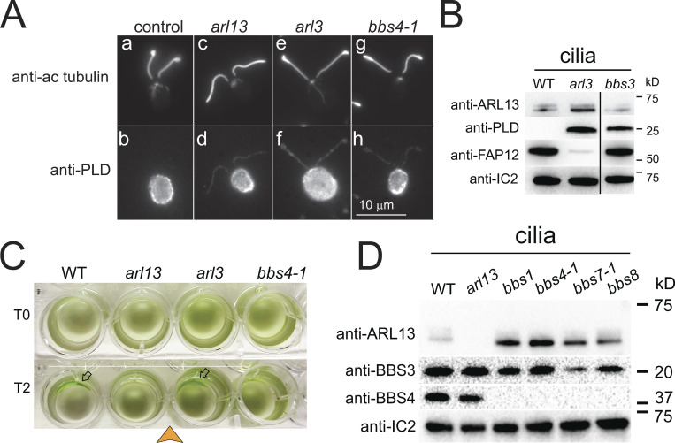
Figure S5.
PLD accumulates along the length of arl13, arl3, and bbs4-1 cilia. (A) Immunofluorescence of methanol (−20°C) treated control (a/b), arl13 (c/d), arl3 (e/f), and bbs4-1 (g/h) cells stained with anti-acetylated tubulin (a, c, e, and g) and anti-PLD (b, d, f, and h). In the cell body, most PLD is located below the plasma membrane. Bar = 10 µm. (B) Western blot of isolated cilia from control (g1, WT), arl3 and bbs3 probed with antibodies against ARL13, PLD, FAP12, and IC2, as a loading control. The black line indicates that an unrelated lane was cropped out; see source file for the uncropped blot. (C) Population phototaxis assay of control (g1, WT), arl13, arl3, and bbs4-1. The direction of light (arrowhead) and time of exposure in minutes are indicated. Arrows, accumulated cells. (D) Western blot of isolated cilia from control (g1, WT), arl13, and bbs mutants probed with antibodies against ARL13, BBS3, BBS4, and, as a loading control, IC2. Source data are available for this figure: SourceData FS5.

