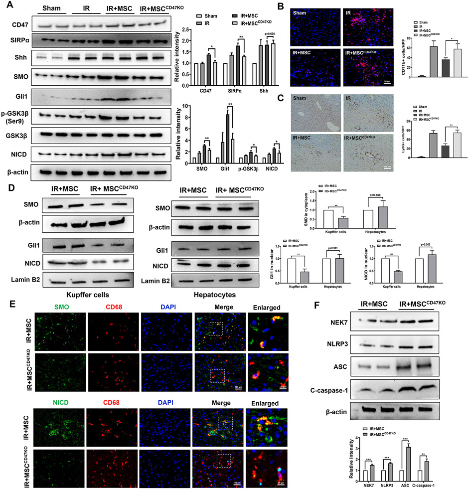
Fig. 2. The CD47-SIRPα interaction activates Hedgehog/SMO/Gli1 pathway, Notch1 signaling, and inhibits NEK7/NLRP3 activation in IR-stressed livers.
WT mice were adoptive transferred MSCs or CD47-deficient MSCs (1x10⁶) 24h prior to liver ischemic insult. (A) Western blot analysis and relative density ratio of CD47, SIRPα, Shh, SMO, Gli1, p-GSK3β, GSK3β, and NICD in ischemic livers. (B) Immunofluorescence staining of CD11b+ macrophages in ischemic livers (n=4-6 mice/group). Quantification of CD11b+ macrophages, Scale bars, 50μm. (C) Immunohistochemistry staining of Ly6G+ neutrophils in ischemic livers (n=4-6 mice/group). Quantification of Ly6G+ neutrophils, Scale bars, 100μm. (D) Western blot analysis and relative density ratio of SMO, Gli1, and NICD in Kupffer cells and hepatocytes. (E) Immunofluorescence staining for SMO or NICD expression in macrophages from the WT liver tissues (n=3-4 samples/group). DAPI was used to visualize nuclei. Scale bars, 100μm and 20μm. (F) Western blot analysis and relative density ratio of NEK7, NLRP3, ASC, and cleaved caspase-1 in ischemic livers. All Western blots represent three experiments, and the data represent the mean±SD. *p<0.05, **p<0.01, ***p<0.001.

