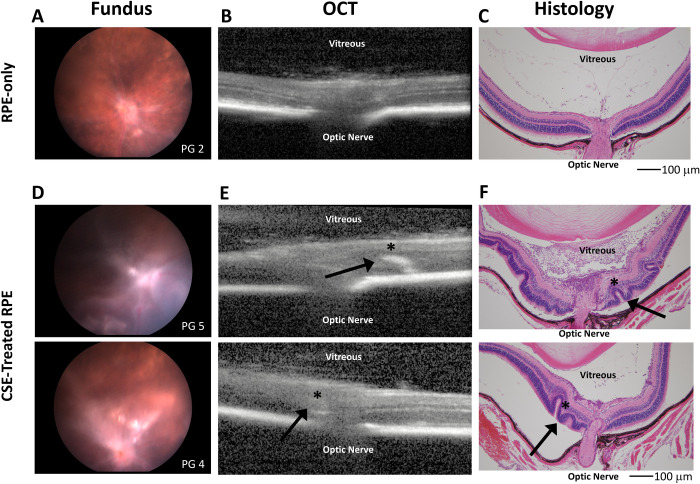
Fig 4. CSE treated RPE cell injection increased retinal folds, retinal detachment and PVR membranes thickness compared to control RPE.
Fundus imaging, OCT, and histology of representative eyes from mice injected intravitreally with control RPE cells (A,B,C) vs. RPE cells treated and resuspended with 0.5% CSE (D,E,F). All images were taken at 4 weeks post-RPE injection. Intravitreal injection of CSE-treated RPE resulted in development of more severe PVR by 4 weeks with evidence of retinal folding, significant areas of detachment, and inflammatory infiltrate. The inflammatory infiltrate is especially prevalent in the vitreous of the top section in F. Arrows and * in E and F highlight retinal folds observed on OCT imaging seen in histologic cross-sections. Scale bars represent 100 microns (μm) in length.

