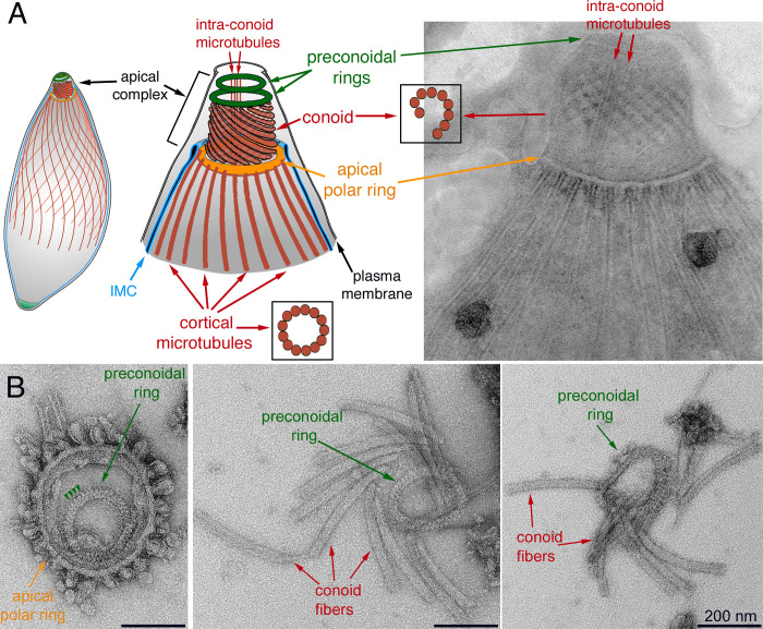
Fig 1. Structure of the apical complex in Toxoplasma.
A. Left and middle: Drawings depicting some of the tubulin-containing structures (red) in T. gondii, including the 22 cortical microtubules, a pair of intra-conoid microtubules, as well as the 14 fibers that make up the conoid. IMC: Inner Membrane Complex. Right: Transmission electron microscopy (TEM) image of a negatively stained TritonX-100 (TX-100) extracted parasite. B. TEM images of the apical parasite cytoskeleton negatively stained after detergent extraction and protease treatment. Left: end-on view of a parasite apical cytoskeleton. Most cortical microtubules and conoid fibers have detached, which allows a clear view of the preconoidal rings lying inside the apical polar ring. Arrowheads indicate the periodic "spikes" in the preconoidal ring. Middle and right: disassembled conoids with attached preconoidal rings.

