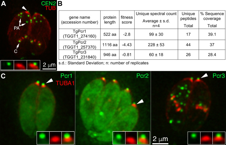
Fig 2. Identification of candidate apical complex proteins via immunoprecipitation using GFP-Trap and eGFP-CEN2 knock-in parasites.
A. Deconvolved wide-field images of intracellular eGFP-CEN2 knock-in parasites [55] expressing mCherryFP tagged tubulin (red). Insets (shown at 2X) include the preconoidal region (arrowhead). PA: peripheral annuli; C: centrioles. B. Table showing the protein length, fitness score, average unique spectral counts, peptide counts, and sequence coverage for TGGT1_274160, TGGT1_257370, and TGGT1_231840, identified by MuDPIT in 4 replicates of immunoprecipitation using GFP-Trap and eGFP-CEN2 knock-in parasites. See S1 Table for the complete list of identified proteins. C. Deconvolved wide-field images of intracellular parasites expressing mCherryFP tagged tubulin (red) and mEmeraldFP (green) tagged Pcr1 (TGGT1_274160), Pcr2 (TGGT1_257370), or Pcr3 (TGGT1_231840) with expression driven by a T. gondii tubulin promoter. As predicted for preconoidal proteins, Pcr1, Pcr2, and Pcr3 are localized to a structure (green, insets) that is apical and smaller in diameter than the conoid (red, insets). Insets (shown at 2X) include the preconoidal region (arrowheads).

