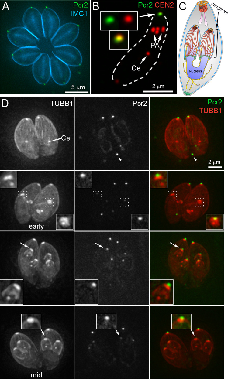
Fig 3. Pcr2 is recruited to the preconoidal region at an early stage of daughter formation.
A. Projection of deconvolved wide-field images of intracellular Pcr2-mNeonGreen 3’ endogenous tag parasites (green) labeled with a mouse anti-IMC1 antibody and a secondary goat anti-mouse Alexa350 antibody (cyan). B. Projection of deconvolved wide-field images of TX-100 extracted extracellular Pcr2-mNeonGreen 3’ endogenous tag parasites (green) labeled with rat anti-CEN2 antibody and a secondary goat anti-rat Cy3 antibody (red). Insets (shown at 2X, contrast enhanced to display weaker signals) include the preconoidal region (arrow). Ce: centrioles. PA: peripheral annuli. C. Drawing depicting a dividing parasite with daughters developing inside the mother cell. For simplification, the cortical microtubules of the mother parasite are not shown. D. Montage showing projections of deconvolved wide-field images of intracellular Pcr2-mNeonGreen 3’ endogenous tag parasites transiently expressing mAppleFP-β1-tubulin (TUBB1, red) from a T. gondii tubulin promoter. The cortical microtubules in the mother parasites are present and clearly seen in the single slices of the 3-D stack, but not clearly visible in these projections due to the decreased contrast in the maximum intensity projection for weaker signals. Pcr2 (green) is recruited to the newly formed apical cytoskeleton as soon as the daughters are detectable. Top row: interphase parasites. Row 2–4: parasites with daughters from early to mid-stage of assembly. Insets (shown at 2X, contrast enhanced to display weaker signals) include the apical region of one of the daughter parasites indicated by arrows. Ce: centrioles. Arrowhead: a Pcr2-mNeonGreen concentration is occasionally seen in the basal region of these parasites.

