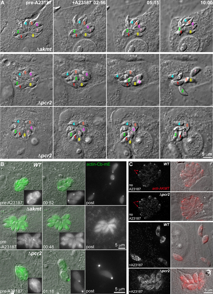
Fig 8. Pcr2 functions differently from another motility regulator, AKMT, and Pcr2 knockout does not block actomyosin activity.
A. Images selected from time-lapse experiments of intracellular Δakmt and Δpcr2 parasites treated with 5 μM A23187. Note that Δakmt parasites, which are largely immobile, maintained their organization within the vacuole after lysis of the host cell. However the position and orientation of the Δpcr2 parasites shifted during the time lapse due to sporadic parasite movement (see S4 Video). B. Actin-chromobody-mEmerald (actin-Cb-mE) distribution before and after A23187 treatment in RHΔhxΔku80 parental (WT), Δakmt and Δpcr2 parasites. The grayscale actin-Cb-mE fluorescence images are projections of the image stack at the corresponding time point. C. Localization of AKMT in the RHΔhxΔku80 parental (WT) and Δpcr2 parasites before and after ~ 5 min 5 μM A23187 treatment. Before exposure to A23187, AKMT is concentrated at the apical end (arrows) of intracellular WT and Δpcr2 parasites. The increase in intra-parasite [Ca2+] caused by A23187 treatment triggers the dispersal of AKMT from the parasite apical end in both the parental and the Δpcr2 parasites. AKMT was labeled by immunofluorescence using a rat anti-AKMT antibody and a secondary goat anti-rat Cy3 antibody. The grayscale anti-AKMT fluorescence images are projections of deconvolved image stacks.

