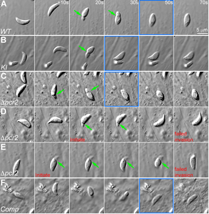
Fig 9. The Δpcr2 parasite moves fitfully during invasion.
Images selected from time-lapse recording of RHΔhxΔku80 (WT, A), mEmeraldFP-Pcr2 knock-in (KI, B), knockout (Δpcr2, C-E), and complemented (Comp, F) parasites in the process of invasion or attempted invasion. D and E show two examples of abortive invasion by the Δpcr2 parasite. The frames where the invasion has completed are marked in blue. Green arrows: constrictions formed during invasion. Also see S8 Video.

