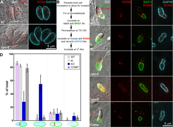
Fig 10. Δpcr2 parasites are defective in assembling the moving junction.
A. Projections of deconvolved wide-field fluorescence images of intracellular mE-Pcr2 KI (KI) and Δpcr2 parasites labeled with a mouse anti-RON4 (red), a rat anti-GAP45 (cyan) and corresponding secondary antibodies. B. Outline of a pulse invasion assay to analyze the assembly of the moving junction (marked by anti-RON4 labeling), and the differential accessibility of the intracellular vs extracellular portion of the invading parasites to antibody labeling of the SAG1 surface antigen. C. DIC and projections of deconvolved wide-field fluorescence images of RHΔhxΔku80 (WT), mEmeraldFP-Pcr2 knock-in (KI), knockout (Δpcr2) and complemented (Comp) parasites, in which SAG1 (green) RON4 (red), and GAP45 (cyan) were labeled by immunofluorescence in the pulse invasion assay described in B. Two predominant patterns are included. D. Quantification of all four SAG1 and RON4 labeling patterns observed in WT, KI, Δpcr2 and complemented (Comp) parasites from three independent biological replicates. Error bars: standard error. * P value <0.05 (unpaired Student’s t-tests), when compared with WT parasites.

