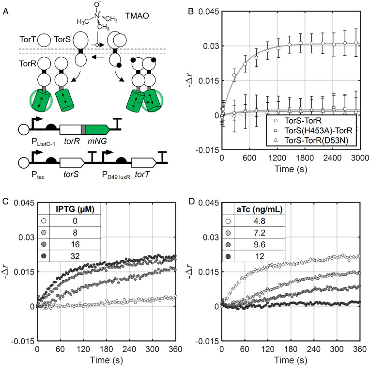
Fig. 2.
Real-time detection of TorR dimerization in E. coli. (A) The mNG-labeled TorSR expression system. TorS contains additional REC and HPt domains C-terminal to the canonical transmitter domain. We omitted these domains from the cartoon schematic because they do not conceptually alter phosphotransfer from TorS to TorR. (B) Change in anisotropy signal for the engineered TorSR system from A and the two negative control systems in response to addition of 1 mM TMAO at time 0. Fit lines are first-order exponential functions (Eq. 2), and fit parameters are given in SI Appendix, Table S4. TorS, TorR-mNG, and TorT were induced with 32 µM IPTG, 4.8 ng/mL aTc, and 1 µM AHL, respectively. (C) Kinetics of TorR-mNG dimerization at different levels of TorS induction with constant TorR-mNG and TorT induction (4.8 ng/mL aTc and 1 µM AHL, respectively). (D) Kinetics of TorR-mNG dimerization at different levels of TorR-mNG induction with constant TorS and TorT induction (32 µM IPTG and 1 µM AHL, respectively). All data points represent the mean of three biological replicates collected on separate days. Error bars in B represent SD. Raw anisotropy values are found in SI Appendix, Table S3.

