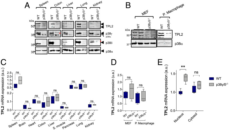
Fig. 1.
p38γ and p38δ regulate TPL2 protein levels. (A) Lysates of the indicated tissues from WT or p38γ/δ−/− mice were immunoblotted with anti-TPL2, -p38α, -p38γ, or -p38δ antibodies. p38α expression was used as the loading control. The red asterisks indicate nonspecific protein bands. (B) MEFs and peritoneal macrophage lysates from WT or p38γ/δ−/− mice were immunoblotted with anti-TPL2 or -p38α. (C) qPCR of TPL2 mRNA from indicated WT or p38γ/δ−/− tissues. Results were normalized to GAPDH mRNA expression. Data show mean ± SEM (n = 3) from one representative experiment of two with similar results. (D) qPCR of TPL2 mRNA from indicated WT or p38γ/δ−/− cells. Results were normalized to GAPDH mRNA expression. (E) qPCR of TPL2 mRNA in cytosolic or nuclear RNA from WT or p38γ/δ−/− MEFs. Results were normalized to GAPDH mRNA expression. Data show mean ± SEM from one representative experiment of two with similar results. ns, not significant; ***P ≤ 0.001 relative to WT.

