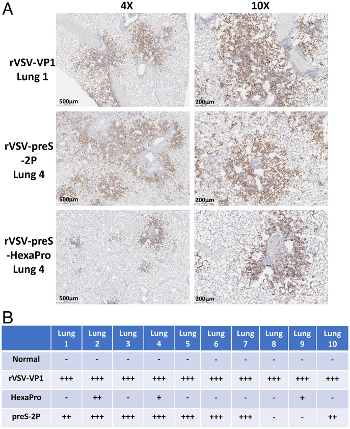
Fig. 12.
Comparison of N antigen expression in lungs of hamsters immunized with rVSV-preS-2P and rVSV-preS-HexaPro after SARS-CoV-2 WA1 challenge. (A) Representative images of immunohistochemistry (IHC) staining of lung sections. Hamsters were euthanized at day 4 after SARS-CoV-2 challenge. Lung sections were stained with anti-SARS-CoV-2 N antibody. Micrographs with 4× and 10× magnification are shown. Scale bars are indicated at the left corner of each image. (B) Summary of IHC staining of 10 hamsters in each group. HexaPro denotes rVSV-preS-HexaPro group and preS-2P denotes rVSV-preS-2P group. “+++” indicates extensive N antigen detected in lung section. “++” indicates moderate amount of N antigen detected. “+” indicates occasional or small amount of N antigen detected. “–” indicates negative for N antigen.

