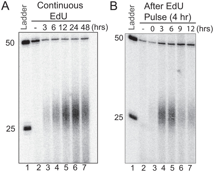
Fig. 4.
Decay kinetics of the EdU-induced excision products in human cells. (A) In vivo excision assay showing EdU excision dynamics. HeLa cells were treated with 10 μM EdU, for 3, 6, 12, 24, and 48 h. Then, the excised oligonucleotides were isolated, immunoprecipitated with anti-TFIIH antibodies, mixed with a 50-mer internal control, 3′ end labeled, and separated on DNA-sequencing gels. (B) In vivo excision assay of EdU pulse labeling and decay kinetics. HeLa cells were pulse labeled for 4 h in medium containing 10 μM EdU. Then, EdU-containing medium was removed and cells were supplied with fresh medium and incubated at 37 °C for 0, 3, 6, 9, and 12 h. Then, the excised oligonucleotides were isolated, immunoprecipitated with anti-TFIIH IP antibodies, and analyzed as in (A).

