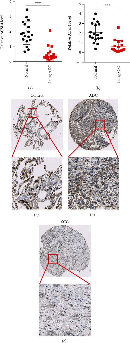
Figure 2.

Compared with normal lung samples, NSCLC tissue samples showed a considerable reduction in ACSL4 expression. (a, b) A qPCR was utilized to examine ACSL4 mRNA expression in 18 pairs of NSCLC and corresponding neighboring normal lung tissues. ACSL4 IHC demonstrated strong staining in normal lung tissues (c); weak staining in NSCLC tissue (d, e). ∗∗∗p < 0.001 vs. control.
