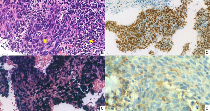
FIGURE 1.

(a) Lymphoepithelial carcinoma (LEC) shows the sheet of poorly differentiated squamous cell carcinoma. The tumor shows a sheet of spindle‐shaped cells with eosinophilic cytoplasm and basophilic nuclei (asterisk) with some lymphocyte infiltrate accompanied by surrounding lymphoplasmacytic stroma (arrow) (hematoxylin–eosin, original magnification ×400). (b) p40 immunohistochemistry in LEC. The tumor cells show diffuse and strong nuclear p40 staining indicating squamous differentiation (original magnification ×400). (c) Epstein–Barr virus (EBV)‐encoded small RNA (EBER) in situ hybridization in LEC. EBER is diffusely positive in the tumor cells (original magnification ×400). (d) Low expression of PD‐L1 (22C3) immunohistochemistry in LEC. The tumor cells show faint incomplete membrane staining of the PD‐L1 staining (original magnification ×600)
