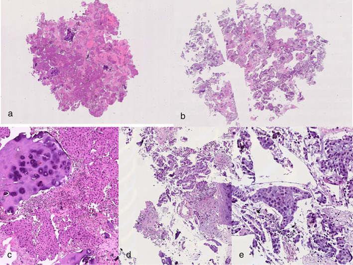
FIGURE 2.

Haematoxilin and eosin (H&E) stained sections (4×) from mediastinal lymph node (a) and pulmonary mass (b), respectively. (c) Cartilage and lymphocytes adjacent to neoplastic component (H&E, 10×). (d) Adenocarcinoma histotype is visible on H&E stained section (10×; E, 20×)
