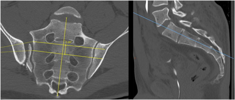
Fig. 2.
(a) Axial reformatted computed tomography (CT) scan showing the angle between the oblique S1 corridor and the sagittal plane, (b) axial reformatted CT scan showing the angle between the transsacral S2 corridor and the sagittal plane, (c) coronal reformatted CT scan showing the angle between the oblique S1 corridor and the sagittal plane, and (d) coronal reformatted CT scan showing the angle between the transsacral S2 corridor and the sagittal plane.


