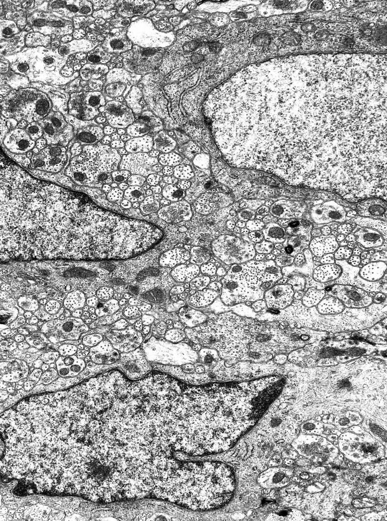
Fig. 2.
A panoramic view of the neuropil by electron microscopy. A nerve cell with nucleus is at top right corner; a nucleated glial cell is half-way along the left border, and another one is at the bottom. The section is transverse longitudinal, with the circular muscle side at the top. If a piece of tissue of this size could be cut again, orthogonally, it would produce more than 130 vertical sections. Width of the microscopic field: 12 µm

