FIGURE 4.

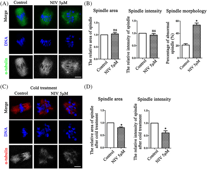
NIV affects microtubule stability in oocyte meiosis. (A) The spindle size and microtubule signals were not affected after NIV exposure compared with the control oocytes, while the spindle morphology of treatment groups was disrupted (Control: n = 45; NIV group: n = 36). Green, α‐tubulin; Blue, DNA; Bar = 10 μm. (B) The relative fluorescence intensity analysis of microtubule intensity and spindle area calculation showed no significant difference between the control and NIV‐treated groups, while the abnormal rate of spindle morphology was much higher after NIV exposure. ns, p > 0.05; *p < 0.05. (C) After 9.5 h culture, the oocytes preformed 5 min cold treatment were used to test the stability of microtubules. After exposure to NIV, the spindle area and microtubule signals were both lower than that in the control groups (Control: n = 33; NIV group: n = 32). Red, α‐tubulin; Blue, DNA; Bar = 10 μm. (D) Fluorescence intensity analysis showed that spindle area and microtubule intensity in the NIV‐treated group were both lower compared with control group. *p < 0.05.
