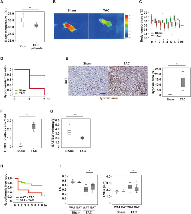
Figure 1.
Left ventricular pressure overload induces dysfunction of brown adipose tissue. (A) Body temperature of control (Con) subjects and patients with congestive heart failure (CHF) (n = 15, 9). (B) Surface body temperature measured by a thermal camera in mice at 5 weeks after sham surgery (Sham) or TAC. (C) Acute cold tolerance test performed in mice at 4 weeks after Sham or TAC with measurement of body temperature in the scapular region (n = 6 and 7, respectively). (D) Hypothermia-free ratio during the acute cold tolerance test in mice at 4 weeks after Sham or TAC with measurement of the intraperitoneal temperature (n = 4, 4). (E) Pimonidazole staining of BAT in mice from (C) performed by the Hypoxyprobe-1 method. The right panel shows quantification of the hypoxic area (n = 4, 8). Scale bar = 50 μm. (F) Quantification of TUNEL-positive cells in BAT from mice at 6 weeks after Sham or TAC (n = 3, 3). (G) Body weight (BW)-adjusted BAT weight in mice prepared as described in Fig. 1C (n = 5, 4). (H) Hypothermia-free ratio during the acute cold tolerance test in mice with WAT or BAT transplantation at 2 weeks after TAC (n = 11, 17). (I) Assessment of cardiac function in mice with WAT or BAT transplantation at 2 weeks after Sham or TAC. FS: fractional shortening (n = 10, 11, 9, 19), LVDs: left-ventricular systolic dimension (n = 10, 11, 9, 19). Data were analysed by the 2-tailed Student’s t-test (A, E, F, G and I), repeated measures followed by Tukey’s multiple comparison test (C), or the log-rank test for Kaplan–Meier method (D, H). *P < 0.05, **P < 0.01. Values are shown as the mean ± s.e.m.

