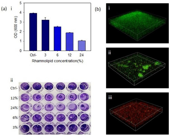
Figure 4.
(a) Investigating biofilm formation by crystal violet staining. (i) Relationship between rhamnolipid concentration and inhibition of MRSA biofilm. (ii) Image of biofilm formed by MRSA bacteria in untreated state and treated with different concentrations of rhamnolipid (3%, 6%, 12%, 24%) with a pH of 7. (b) Confocal microscope analysis of MRSA biofilms. (i) Untreated biofilms, and (ii, iii) treated biofilm with concentrations of 6% and 24%, respectively.

