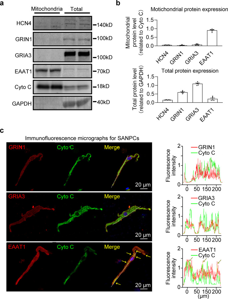
Fig. 6. EAAT1 was abundantly expressed in rat SANPC mitochondria.
a Western blot analysis confirmed that EAAT1 was abundantly expressed in SANPC mitochondria. b Pooled data from a (n = 5 per groups). c Immunofluorescence staining with anti-EAAT1 antibody, anti-GRIA3 antibody, anti-GRIN1 antibody and anti-Cytochrome C antibody (mitochondrial marker) in isolated SANPCs (left); line plot profiles of the fluorescence along the white dotted line on the left (right).

