
Figure 5.
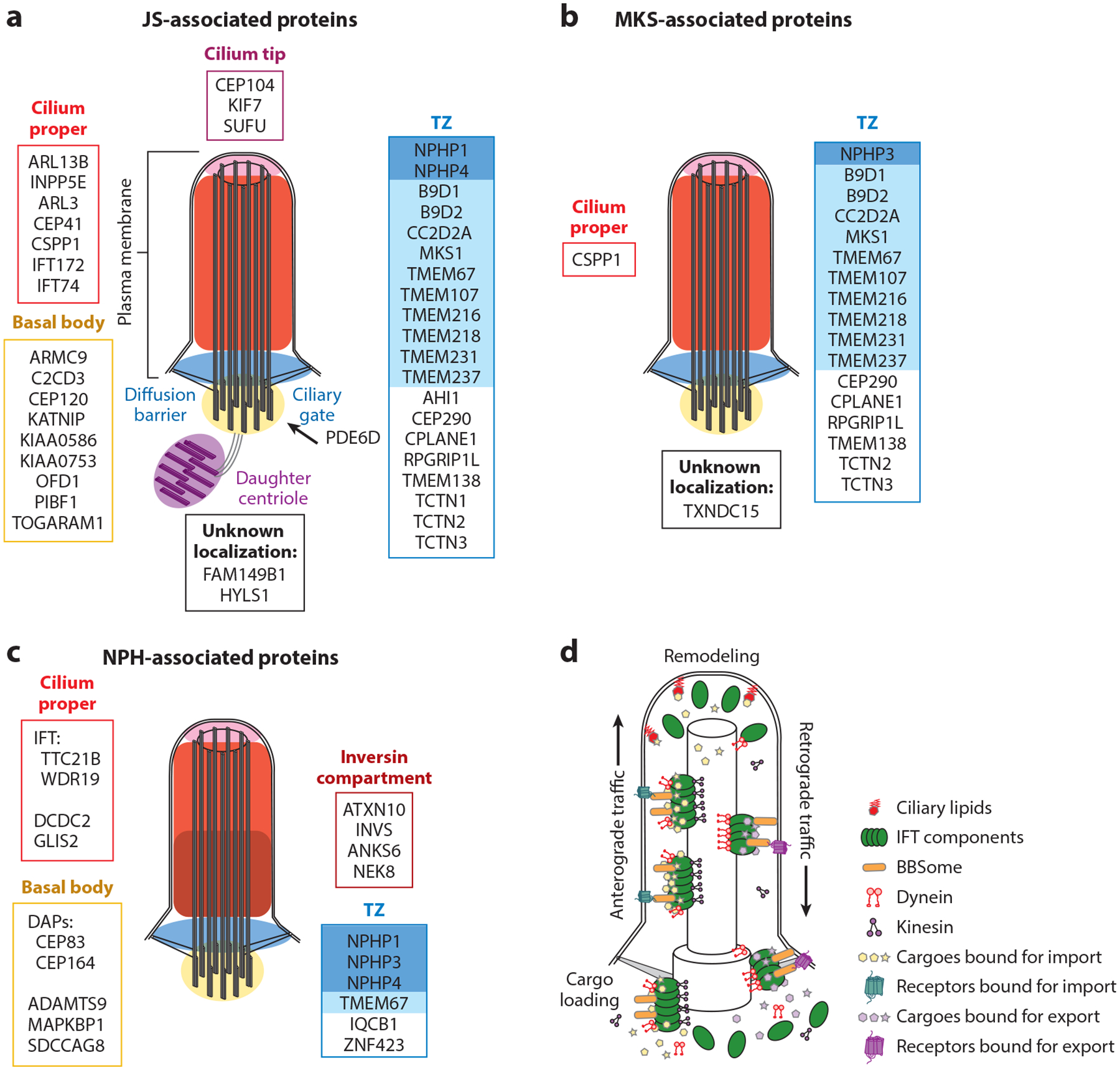
JS-MKS-NPH protein localization and IFT. (a) JS-associated proteins grouped by their localization in steady-state cilia. The TZ is subdivided into the NPHP module (dark blue) and the MKS module (light blue). The ciliary membrane is continuous with the plasma membrane. (b) MKS-associated proteins grouped by their localization in steady-state cilia. (c) NPH-associated proteins grouped by their localization in steady-state cilia. (d) Transport of ciliary proteins by IFT components (green). The BBSome (orange) traffics receptors. Ciliary lipids (red) capture and retain specific proteins. Abbreviations: BBSome, protein complex involved in Bardet–Biedl syndrome; DAP, distal appendage; IFT, intraflagellar transport; JS, Joubert syndrome; MKS, Meckel syndrome; NPH, nephronophthisis; TZ, transition zone.