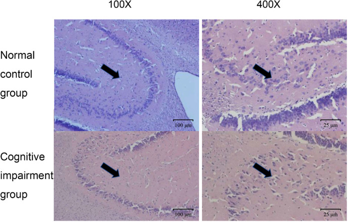
Figure 2.
Comparison on pathological changes of hippocampal neurons from rats in the normal control group and the cognitive impairment group. Representative images of HE staining results from the normal control group and the cognitive impairment group. The hippocampal neurons were clear in structure, normal in size, and stained light blue; while those in the cognitive impairment group were not arranged orderly, largely degenerated, and necrotic. Magnification, 100 × and 400 ×, as indicated.

