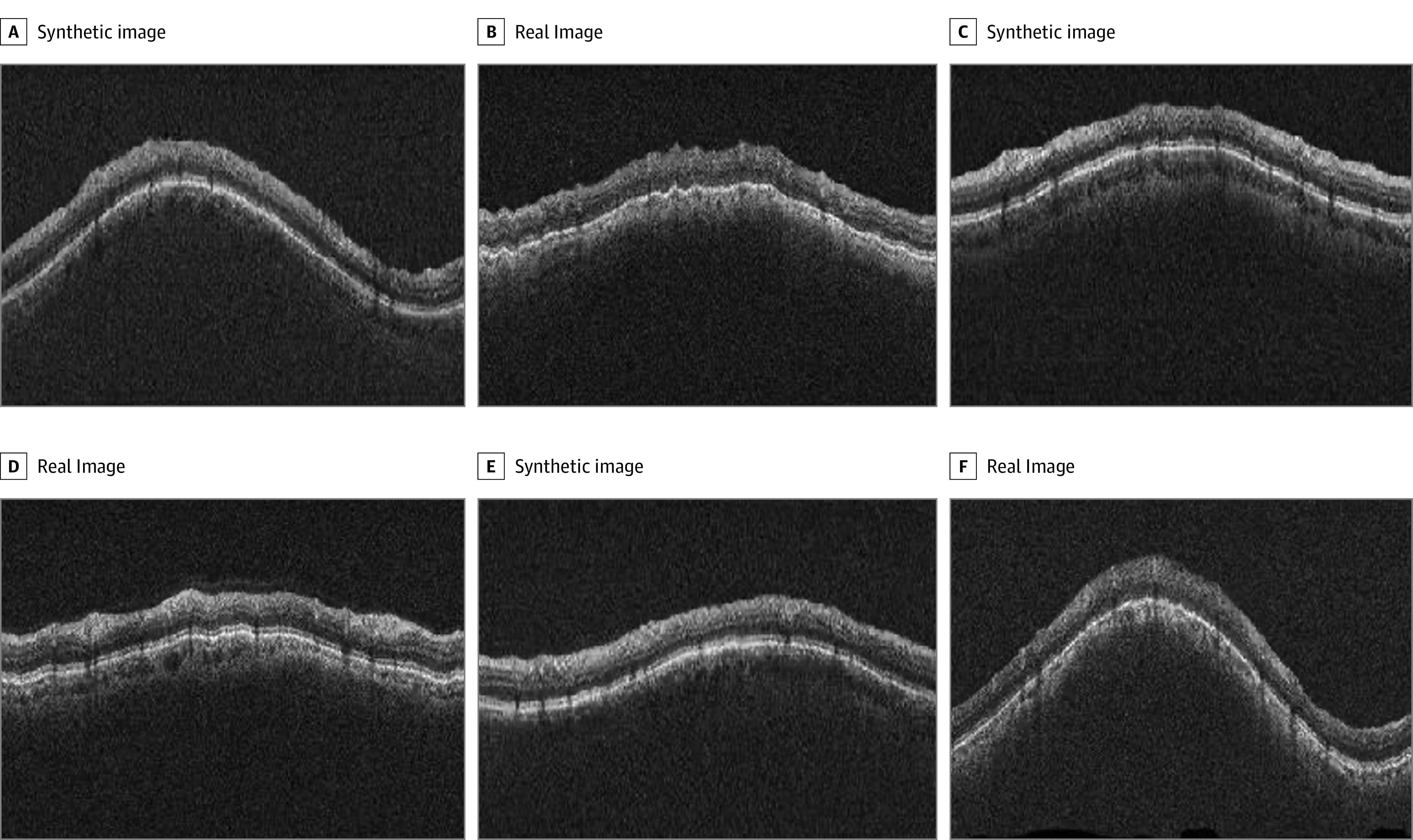
Figure. Circumpapillary Optical Coherence Tomography (OCT) Images of Real and Synthetic Glaucomatous Eyes.

Examples of images of circumpapillary OCT images from real participants and synthetically generated circumpapillary OCT images from the generative adversarial network (GAN) model for glaucomatous eyes. Circumpapillary OCT images of real glaucomatous eyes are located in panels B, D, and E. All other images were synthetically generated from the GAN model for glaucomatous eyes.
